At a Glance
- A group of UVA cardiologists, radiologists and biomedical engineers are collaborating to expand on and develop new imaging techniques for cardiac patients.
- UVA imaging experts are using MRI of the heart to make procedures for heart rhythm problems and heart failure problems more effective.
- Medtronic’s CardioInsight™ 3-D body surface electrocardiographic mapping system offers cardiologists a noninvasive alternative that can lead to a more comprehensive diagnosis during catheter ablation and pacemaker/implantable cardioverter defibrillator (ICD) procedures.
- UVA imaging experts pioneered a way to make transesophageal echocardiogram unnecessary for select patients undergoing ablation for atrial fibrillation.
- UVA is making MRI available to patients with pacemakers and defibrillators.
- UVA has recently adopted the CardioMEMS™ HF System to better manage patients with pulmonary hypertension.
Over the last decade, the power, versatility and variety of techniques available for imaging patients with heart failure, arrhythmias and pulmonary hypertension have expanded dramatically. The challenge now for medical centers is to orchestrate the application of these techniques to provide the best possible care for patients. This year, UVA has assembled a team of cardiologists, radiologists and biomedical engineers to not only optimize the use of these techniques for patient care, but also to advance the field by developing new and more effective ways to deploy them.
“Our goal is to stay out ahead of the curve so that we are prepared to make the best possible use of new developments, while pioneering new techniques ourselves,” says Kenneth Bilchick, MD, MS, director of the UVA Program for Advanced Imaging in Cardiac Electrophysiology and Heart Failure.
Bilchick points to the hardware and software innovations that have expanded the utility of such established techniques as magnetic resonance imaging (MRI), cardiac computed tomography (CT), positron emission tomography (PET), single-photon emission computed tomography (SPECT) and echocardiography. At the same time, there have been major advances in 3-D electroanatomic mapping (EAM) systems used during arrhythmia procedures to identify the target areas for ablation. “By combining our expertise, our group hopes to more fully realize the potential of these innovations,” Bilchick says.
Pioneering New Imaging Techniques for Catheter Ablation of Heart Rhythm Disorders
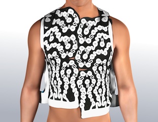
UVA is investigating the use of Medtronic’s CardioInsight™ 3-D body surface electrocardiographic mapping system. This system will offer cardiologists a noninvasive alternative that can lead to a more comprehensive diagnosis during catheter ablation procedures designed to provide relief from symptoms caused by ventricular tachycardia, atrial fibrillation and atrial flutter. The system may also help make complex device procedures for patients in need of cardiac resynchronization therapy (CRT) more effective.
The multielectrode CardioInsight mapping vest captures electrical signals from the body surface and combines them with anatomical data generated from a flash CT image to produce a real-time, electrical 3-D map of the heart. “Although our current EAM technology during the procedure is excellent, this technology may give us an additional advantage by allowing us to target key anatomical areas for ablation before the start of arrhythmia procedures and may also help us identify optimal pacing strategies for patients receiving certain types of pacemakers and defibrillators,” says Bilchick.
Just how instrumental this technology will be is yet to be determined. “We are actively involved in finding the best way to use this new modality,” Bilchick says. Heart rhythm specialists from the UVA Advanced Imaging group — including Bilchick; John Ferguson, MD, , MBChB; J. Michael Mangrum, MD; Pamela Mason, MD; Andrew Darby, MD; and Rohit Malhotra, MD — currently have several approved research protocols under way to study how cardiologists can achieve optimal results by using the CardioInsight vests to complement the standard ablation and device procedures.
Making Atrial Fibrillation Procedures Easier for Patients
Members of the UVA advanced imaging program are also working to make it less stressful for patients to undergo treatment for atrial fibrillation and flutter. Previously, on the day before their ablation, patients underwent both a cardiac CT exam to evaluate pulmonary vein anatomy and a transesophageal echocardiogram (TEE) to evaluate the left atrial appendage for blood clots (thrombus). This required patients to undergo an additional invasive procedure and remain fasting for a good part of the day prior to their ablation.
To streamline the process and maximize patient comfort, Bilchick and colleague Michael Salerno, MD, PhD, pioneered a way to integrate cardiac CT angiography acquired for pulmonary vein anatomy into the care of these patients in order to rule out left atrial appendage thrombus. Patrick Norton, MD, and Klaus Hagspiel, MD, of the UVA Department of Radiology and Medical Imaging were key contributors; nursing staff, including Elaine Knight, RN; Cherie Parks, RN; and Susan Gionakos, RN, were also instrumental in making the project successful.
Their approach, using delayed imaging after contrast administration, minimizes the false positives, and rules out thrombus in many patients with a high degree of accuracy, according to outcomes recently published in the January 2016 edition of Heart Rhythm Journal. Now patients can come in for a cardiac CT and only undergo TEE if there is an equivocal or positive result. Developing this protocol required coordination among electrophysiologists, radiologists, nurses and the Heart Rhythm Quality and Safety Team. “By working together across departments,” Bilchick says, “we raised patient satisfaction, improved safety, increased efficiency and decreased cost.”
Developing Techniques for Treating Pulmonary Hypertension and Heart Failure
Collaborations among members of the advanced imaging program have also led to promising advances in imaging for patients with pulmonary hypertension. By working together with biomedical engineering investigators Salerno and Frederick Epstein, PhD, the UVA team successfully adapted an MRI method previously used to detect diffuse fibrosis in the left ventricle for use with the thinner right ventricle, the chamber often damaged by pulmonary hypertension. This information was then combined with MRI information on RV function, size and morphology to provide a comprehensive assessment for these patients.
In addition, thanks to an effort led by Jamie L.W. Kennedy, MD, and Sula Mazimba, MD, MPH, UVA has recently been offering the CardioMEMS™ HF System from St. Jude Medical to help patients with heart failure, some of whom also have pulmonary hypertension. This implantable pulmonary pressure monitor can provide highly useful information on right-sided heart pressures without the requirement for additional heart catheterization procedures. The use of MRI evaluation of the structure, function, tissue characteristics of the right ventricle, as well as intracardiac monitors of pulmonary pressures, is helping UVA cardiologists guide therapies and monitor their effectiveness.
Making Cardiac Resynchronization Therapy More Effective
MRI offers one of the best ways to optimize CRT both before and after the procedure. The members of the advanced imaging program have harnessed the power of MRI for this purpose by developing methods to image optimal pacing sites prior to the procedure and being among the first to gain access to MRI-conditional CRT devices to facilitate post-device cardiac MRIs in these patients. They have also developed a protocol for safely offering MRIs to patients with the older CRT devices.
Also of note: This team developed a new way of imaging the heart that minimizes artifacts generated by pacemakers and ICDs, which has resulted in high-quality MRI images even in patients with these devices. This is especially useful for patients with ventricular tachycardia who stand to benefit from a detailed assessment of myocardial scar by MRI prior to catheter ablation. UVA’s new strategy combines wideband late gadolinium enhancement (LGE) MRI, which modifies the bandwidth of image acquisition and blocks frequencies that cause artifacts, with a strain imaging sequence developed at UVA. The result is excellent MRI images for cardiac function, scar and strain.
“Taking a systematic approach to evaluating innovations in cardiac imaging and pinpointing areas where we can make a contribution is one way that we can deliver substantial improvements in patient care,” Bilchick said. “This multidisciplinary program is doing just that.”
If you would like to receive a visit from one of the UVA Heart & Vascular team members, contact the UVA Physician Relations team.