When your patient learns her mammogram looks suspicious, it’s hard for her not to imagine the worst. She may face the discomfort of a biopsy, as well as the anxiety of wondering whether she will be among the approximately 25 percent of patients whose biopsies prove positive.
Mark B. Williams, PhD, has devoted the past decade to developing a new scanner that he hopes will give women and their physicians better diagnostic information from the start. His three-dimensional, dual-modality tomosynthesis (3-D DMT) scanner offers a number of advantages. It combines two complementary breast imaging techniques that taken together could identify smaller masses and more accurately pinpoint their locations, distinguish between cancerous and harmless lesions and provide quantitative information about these lesions if they are cancerous.
“The scanner could lead to earlier treatment for women with breast cancer and spare other women the pain and anxiety of undergoing a biopsy,” Williams says. To determine the predictive value of images produced by the 3-D DMT, Williams has launched a clinical trial now enrolling at UVA.
Combining Form and Function
Traditional mammograms use X-rays to generate information about the anatomical structure of a lesion. One of the advantages of the 3-D DMT is that it combines X-ray images with molecular breast images, which shed light on the functional status of breast tissue. Williams uses an FDA-approved tracer that moves through the body and accumulates preferentially in cells with a high metabolic rate and a large blood supply. Both of these states are characteristic of malignant breast cancer cells. The 3-D DMT scanner runs the X-ray and molecular scans sequentially, obtaining both types of images with the breast in the same immobilized position. “This hybrid imaging approach permits the anatomic and functional images to be accurately combined, and gives radiologists the ability to crosscheck the abnormalities they spot using one modality with information from the other,” William says.
Adding a New Dimension
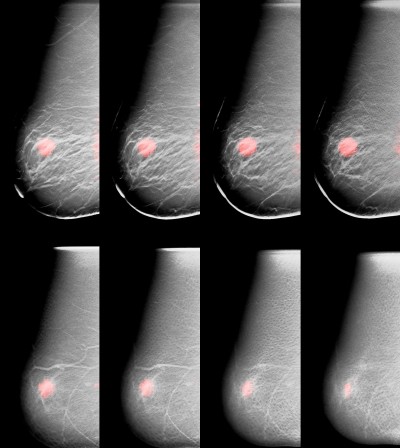
Looking at a photograph, it becomes apparent that information is lost when three-dimensional reality is compressed into two dimensions. This is due to a phenomenon called superimposition. Objects in the foreground cover objects in the background, and background objects often make foreground objects difficult to perceive. Superimposition makes it difficult for radiologists to interpret two-dimensional mammograms, especially those of the 40 to 50 percent of women whose breasts contain a large fraction of fibrous and glandular tissue, so-called “dense” breasts. This type of tissue, while normal and healthy, can mask abnormalities in mammograms, making them undetectable.
Williams’ solution is to employ a technique called tomosynthesis to separate the superimposed elements and produce three-dimensional versions of both his X-ray and molecular scans. In dual modality tomosynthesis, the DMT scanner takes a series of X-ray images of the breast at slightly different viewing directions across an arc that ranges from 15 to 45 degrees. This is immediately followed by a series of images of the tracer over a range of viewing angles. The images of each modality are combined mathematically to construct a series of planes (slices) that a radiologist can examine individually.
This three-dimensional view is clearer: smaller and subtle abnormalities are more readily apparent because they are not embedded in background information. Having both the X-ray slices and their corresponding tracer slices available permits the radiologist to simultaneously assess both anatomical and physiological information about an abnormality and characterize it more accurately.
Comparing Images
Williams and his team have launched a trial, funded by the NIH and Susan G. Komen Foundation, to determine if 3-D DMT is better than mammography in identifying small abnormalities and differentiating benign tumors from cancerous ones. This trial is only available at UVA. The study will involve:
- An injection of a small amount of an FDA-approved radioactive imaging tracer
- Positioning on the 3-D DMT scanner, which requires less breast compression than a mammogram
- X-ray tomosynthesis (X-ray exposure is the same as that of a mammogram)
- Molecular breast tomosynthesis
- Repeat of the procedure on the subject’s other breast
As part of the trial, radiologists will read the X-ray and molecular tomosynthesis images, both individually and in combination, and their findings will be compared to the biopsy results to determine the predictive value of 3-D DMT.
Taking a Look Ahead
If 3-D DMT represents an improvement over traditional mammography, it could transform breast cancer diagnosis, but it also could also help improve treatment. For instance, oncologists could use it to track the effects of radiation or chemotherapy on a tumor. As the FDA approves more targeted tracers, 3-D DMT could produce additional information that would help radiologists more accurately characterize malignant tumors and assist oncologists in their selection of appropriate treatments.
Study Eligibility
The study is enrolling 200 women who meet the following criteria. Participants should:
- Be between 18 and 80 years of age.
- Have had a mammogram in the last 30 days
- Be scheduled for a biopsy
The entire study session will take approximately an hour, and volunteers will receive $100 in compensation for participating.
To find out if your patient is eligible to participate in this study of 3-D dual-modality breast imaging (IRB-HSR# 8825), please contact Andrew Polemi at 303.229.3341 or amp3as@virginia.edu, or Mark B. Williams at 434.953.6083 or mbwilliams@virginia.edu.