In 1831, Michael Faraday discovered that a changing magnetic field induces an electric current in nearby wires. While Faraday is likely an unknown figure to most, this fundamental principle of electromagnetism is significant for patients with pacemaker and implantable cardioverter defibrillator (ICD) systems.
Magnetic resonance imaging (MRI) uses strong magnetic fields, and thus has been off-limits to patients with ICDs in most situations for many years; there is the potential for the wires connected to the heart to overheat and for the device to become damaged. Often MRI is the best — and perhaps the only — imaging modality available to diagnose conditions such as brain tumors, strokes, spine disorders, knee/shoulder injuries, various disorders of the abdominal organs and cardiac viability after a heart attack, so these patients have been at a disadvantage.
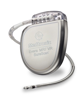
MR-conditional pacemakers have been approved and on the market for the past several years; however, many patients with ICDs have still not been able to get MRIs as the standard of care in recent years. Fortunately, this all changed in September with the introduction of the Medtronic Evera MRI™ SureScan™ ICD system. Now the next generation of ICD patients will be able to have MRIs as standard of care when they need them. UVA was the first medical center in Virginia to implant the Evera system, which complements the Medtronic Advisa MRI pacemakers.
“MR-conditional ICDs allow patients who need these lifesaving devices to have this important diagnostic imaging test,” says electrophysiologist Kenneth Bilchick, MD, who coordinates a UVA program for patients with pacemakers and ICDs who also need MRIs. “We’re committed to making the latest innovations in the field available to patients with heart rhythm disorders as quickly as possible.”
Opening the Door for Greater Access to MRI
Because of the increasing number of patients with ICDs and the increasing value of MRI, researchers have been eager to overcome this hurdle and have studied closely the impact of MRIs on these devices. In fact, while at Johns Hopkins, Bilchick was part of the group that performed the early MRI safety studies for standard cardiac implantable electronic devices, which included validation of a monitoring protocol allowing physicians to provide MRIs for selected patients with these devices.
This protocol has now been implemented at UVA for the past several years in order to provide MRIs for selected patients with standard pacemakers and ICDs. “Even with the new MR-conditional devices, many patients still have standard devices that cannot be easily converted to an MR-conditional system,” explains Bilchick. “The rationale for this protocol is supported by FDA statements to the effect that it is reasonable to perform MRIs in certain patients with standard devices not previously studied for routine MRIs when the benefits are determined to outweigh the risks, and the results are tracked in a registry.”
According the current UVA protocol for these patients, a carefully monitored and supervised scan may be performed at UVA with device reprogramming before and after the scan if all of the following conditions are met:
- Physicians determine that no other imaging modality can provide an adequate answer to the clinical question of interest.
- The patient provides informed consent.
- Certain other device-related criteria are met, such as implantation at least six weeks prior to the MRI date.
After the MRI scan, the results are entered into a registry at UVA, which is among a small number of medical centers that offer this protocol.
While this protocol has been a beneficial development for many, those patients who do not qualify and those patients who do not have access to a center like UVA that offers such a protocol have still been without good options. That’s where Evera comes in. “Even patients with ICDs who are excluded from this protocol can now have an MRI as standard of care not just at UVA, but at most hospitals with MRI and electrophysiology services,” Bilchick says.
A Commitment to Cutting-Edge Care
UVA’s is committed to making the full benefits of MRIs and advanced implantable cardiac devices available to patients with heart rhythm disorders and heart failure, especially because research has shown that implanting ICDs in heart failure patients before they develop arrhythmias saves lives.
Additional examples of UVA’s efforts to push the boundaries of device therapy include a pioneering research program that helps realize the full potential of cardiac resynchronization therapy (CRT) defibrillators, which can both improve heart function and provide protection again sudden cardiac arrest.
Researchers at UVA are also using a novel MRI technique called cine DENSE to image complex regional mechanics in the heart. This technique was developed by UVA Biomedical Engineering Professor Frederick Epstein, PhD, and Bilchick and Epstein have worked together to apply this technique effectively in patients with heart failure. “It gives electrophysiologists the information they need to optimize placement of the pacing leads to maximize heart function and improve associated heart failure symptoms,” says Bilchick. “They can also better identify those patients who will benefit most from cardiac resynchronization therapy.”
“As an academic medical center, we have both the intellectual resources and the responsibility not only to make the latest devices available as they are introduced, but also to pioneer techniques that make them even more effective,” Bilchick says.
Learn more about the devices currently available at UVA for arrhythmia and heart failure.