Recent advancements in pediatric airway surgery have focused on replacing extensive open surgeries with endoscopic techniques. These newer surgeries allow children to recover quickly and avoid prolonged pain, intubation, and tracheostomy.
An early adopter of these endoscopic techniques, UVA Children's helps children with rare pediatric airway problems avoid lengthy, painful recoveries.
Using newer techniques like endoscopic electrocautery devices, airway balloons, Fogarty catheters, lasers, and coblation wands, in combination with increasingly small telescopes (as small as 1.9 mm), UVA Children’s offers more options for children with complex airway problems.
Our multidisciplinary team includes pediatric otolaryngologists, pulmonologists, intensivists, anesthesiologists, speech-language pathologists, surgeons, and gastroenterologists.
Tracheosophageal Fistula Repair
Pediatric surgeons treat most congenital tracheosophageal fistula with open techniques that require a neck dissection or thoracotomy. These put children at risk for prolonged pain, prolonged intubation, injury to vascular structures, injury to the recurrent laryngeal nerve, seroma, and hematoma.
Depending on the size and location of the fistula, UVA Children’s surgeons are able to treat these with endoscopic techniques. “This reduces average length of hospital stay from 7-10 days to 1 overnight stay,” explains UVA Children’s ENT surgeon Ariana Greenwell, MD. “Additionally, children do not require a neck dissection or, potentially, a thoracotomy incision.”
Risks of the endoscopic approach are similar to an open approach, perhaps with a slightly higher recurrence rate. “There have not yet been enough cases described or any head-to-head comparison,” Greenwell says.
Our surgeons perform a dynamic rigid bronchoscopy and pass a urethral stent through the tracheal portion of the fistula. They then pass a telescope into the esophagus to visualize entrance into the esophagus. They use the urethral stent to measure the length of the tract. Surgeons confirm the tract via intraoperative fluoroscopy.
They then use an appropriately sized electrocautery (4-5mm) to cauterize the length of the tract. They typically do this while spontaneously ventilating the child, without using an endotracheal tube. They schedule a second-look procedure and complete additional cauterization as needed.
Piriform Sinus Tract/Fourth Branchial Cleft Sinus Tracts
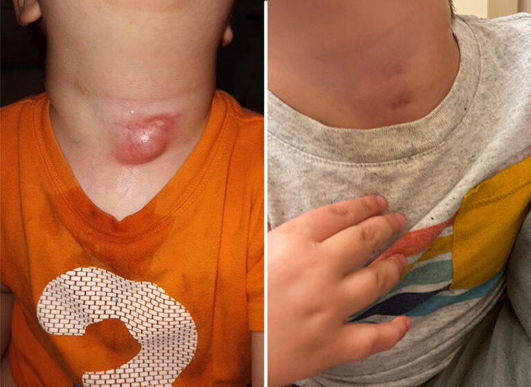
Third and fourth branchial cleft sinus tracts, or piriform sinus tracts, are a rare congenital anomaly representing approximately 2% of all branchial anomalies. They typically present with an anterior neck abscess at the level of the thyroid gland. They can be misdiagnosed as infected thyroglossal duct cysts. Imaging will reveal involvement of the thyroid gland, and they typically present on the left side of the neck.
Historically, surgical excision was the mainstay of treatment. This involved excision of the tract and possible hemithyroidectomy. Risks included external scarring, damage to the recurrent laryngeal nerve, hypocalcemia related to devascularization or resection of the parathyroid glands, and hypothyroidism, which occurs at a higher risk in the pediatric population compared to adults.
At UVA Children’s, we offer an endoscopic treatment approach. Surgeons use suspension microlaryngoscopy to evaluate the piriform sinus, the typical internal opening for these fistulae. We keep the patient spontaneously breathing through the procedure, oftentimes not even requiring placement of an endotracheal tube. Using electrocautery, we cauterize the tract.
Though these have the potential to recur following endoscopic treatment, many parents opt for endoscopic treatment because it is minimally invasive. The procedure also carries a risk of injury to the recurrent laryngeal nerve due to thermal injury. The risk is still lower than with open surgery, but we mitigate it by using low-wattage cautery settings. We observe patients overnight to monitor for any airway edema, and patients typically are discharged home on postoperative day 1 with a regular diet.